Several million tons of plastic waste end up in the oceans every year and degrade into microplastics. These plastic particles, which are only a few micro- or nanometers in size, find their way into the food chain via fish or mussels and therefore end up in the human digestive system. "The health risks posed by microplastics still need to be assessed," says Prof. Dr. Harald Giessen, Head of the 4th Physics Institute and the Stuttgart Center of Photonics Engineering at the University of Stuttgart. "The prerequisite for this is the detection of micro- and nanoplastics in the food chain."
Spontaneous Raman spectroscopy is already being used to detect microplastics in drinking water. This method is based on the fact that molecules exhibit distinct natural vibrational modes. "The laser microscope is used to measure wavelength shifts in the scattered light to identify and distinguish different chemical compounds," says Dr. Moritz Floess, research associate at the 4th Physics Institute. "Unfortunately, this process is very slow. In order to collect larger amounts of data - for example, to quantify the contamination of salmon in the Atlantic with microplastics - much faster imaging is required."
Physicists and neurobiologists conduct joint research
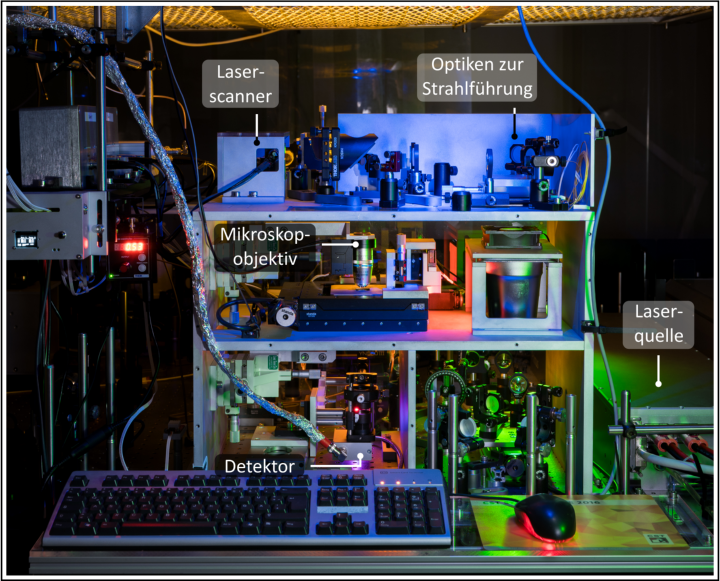
Together with colleagues from the 4th Physics Institute and the Institute of Biomaterials and Biomolecular Systems, Moritz Floess has published a paper in the renowned scientific journal Biomedical Optics Express. The researchers present an approach for the detection of microplastics in fish tissue that could enable big data surveys. The interdisciplinary research team employed stimulated Raman scattering (SRS) instead of spontaneous Raman spectroscopy: "We use two laser beams with different wavelengths. The wavelength detuning is precisely matched to the energy difference associated with the molecular vibration we intend to excite. Therefore, we can define exactly what type of molecule we are looking for and scan the sample pixel by pixel. Thanks to the significantly higher signal level provided by the SRS process, images can be acquired within around 30 seconds compared to about 30 minutes using spontaneous Raman spectroscopy."

Parameter studies with fish from the supermarket
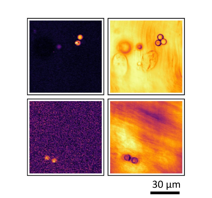
SRS microscopy requires a high level of expertise in the field of laser microscopy and has therefore so far only been used in highly specialized research laboratories. "In our study, we were able to demonstrate for the first time that microplastics can be detected in the muscle tissue of fish using SRS. In particular, we quantified the detection limits: How deeply can we penetrate the tissue with this method? What is the smallest detectable particles size? We are operating in the 100-nanometer range here," says Moritz Floess.
When designing the study, the team decided against experiments with live fish. "We went to a local fish vendor. Professor Ingrid Ehrlich, the neurobiologist on the research team, provided thin slices of muscle tissue, and then we prepared them with microplastic particles. In this way, we created a controlled environment for our parameter studies."

Huge potential for biomedical research
"The potential of SRS does not only lie in rapid imaging. The process also allows to differentiate between different types of microplastics such as polymethyl methacrylate or polystyrene directly under the microscope," says Giessen. In follow-up projects, he and his team plan to track the path of microplastics in the digestive system.
And it's not just microplastics that can be detected using SRS: In the future, the method could also be used in cancer diagnostics. “Research in these areas requires highly specialized laser technologies, which we develop here at the 4th Physics Institute. This requires close interdisciplinary collaboration between biomedicine and physics. Therefore, we are ideally situated in Stuttgart in this respect."
The unique research profile of the University of Stuttgart is characterized by the guiding principle of networked disciplines, known as the "Stuttgart Way". Moritz Floess, a junior researcher who also completed his Bachelor's and Master's degrees at the 4th Physics Institute, recognizes the benefits of the "Stuttgart Way" for his professional growth. "The interdisciplinary project work teaches you to look beyond your own 'science bubble'," he notes. The SRS study was a part of Floess' doctoral thesis. "SRS systems are highly complex and require various specialized components, including laser optics, electronics, measurement technology, microscopy, and programming. It was very valuable for my doctoral thesis that the institute has expertise and support in all these areas."
Terra incognita enables pioneering research in uncharted future research areas
The project was funded by the "Terra incognita" program of the University of Stuttgart. "Terra incognita" was launched in 2019 to identify promising fields of research for the future. Scientists should be encouraged to explore risky and original project ideas, aiming to achieve new research results by taking new approaches. "The program also encourages unconventional research ideas and provides an ideal framework for developing interdisciplinary connections," says Professor Giessen.
Original publication:
Floess, M. Fagotto-Kaufmann, A. Gall, T. Steinle, I. Ehrlich, and H. Giessen, "Limits of the detection of microplastics in fish tissue using stimulated Raman scattering microscopy," Biomed. Opt. Express 15, 1528–1539 (2024).

Lena Jauernig
Editor Research / Early Career Researchers

Moritz Floess
Dr.Stimulated Raman scattering microscopy


